Their DNA is not enclosed within a membrane. Which drawing in Figure 41 possesses an axial filament.
Solved Figure 2 D Which Drawing In Figure 2 Is Chegg Com
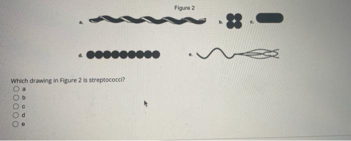
Which drawing in the figure is streptococci.
. Which drawing in the figure is streptococci. Similar to viridans streptococci. They are nonmotile and non-spore forming.
B e a d c Which drawing in the figure possesses an axial filament. Individual cocci are spherical 05-1 µ in diameter and arranged in chains Fig. Which drawing in Figure 41 is a bacillus.
PowerPoint Win Mac compatible. Tap again to see term. The chains are longer in liquid than in solid media.
The Correct Answer is OOOOOOOOOO Reason Explained OOOOOOOOOO is correct for Which drawing in Figure 41 is streptococci. A b c d e d How do spirochetes and spirilla differ. They lack a plasma membrane.
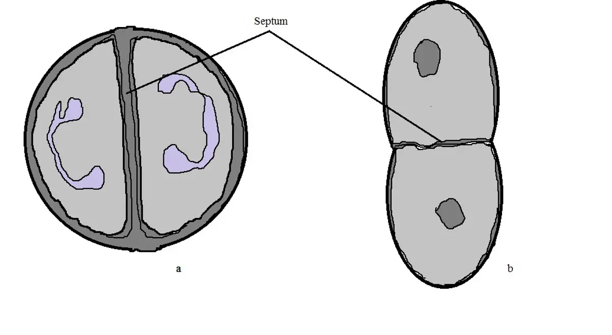
Question 51 Not yet graded 5 pts Bonus Question. Beta refers to complete clearing figure 5 and gamma means there is no lysis. As cellular division of Streptococcus spp.
Streptococci are coccoid bacterial cells microscopically and stain purple Gram-positive when Gram staining technique is applied. In Figure 43 which diagram of a cell wall is a gram-negative cell wall. Tap card to see definition.
Click card to see definition. Streptococci illustration figure drawing diagram image. Pyogenes on different culture media.
If you have any special questions you can comment to ask us. Which microscope achieves the highest magnification and greatest resolution. Occurs along a single axis or plane these bacteria grow in pairs or chains.
They reproduce by binary fission. E b a c Which of the following statements is INCORRECT regarding prokaryotic cells. A microscope 30-50X or a 3X hand lens can also be a useful tool in differentiating pneumococci from viridans streptococci.
Which drawing in Figure 41 is streptococci. Alpha refers to partial hemolysis with a green coloration from production of an unidentified product of hemoglobin seen around the colonies. Which drawing in Figure 41 is streptococci.
Spirochetes and spirilla are basically the same organisms and the terms can be used interchangeably. Spirilla are found in chains of cells whereas spirochetes exist as individual cells. Which drawing in Figure 41 is streptococci.
Which of the following structures is NOT found in prokaryotic cells. There are several differences between prokaryotic and eukaryotic cells. We hope you get all your answers here.
Older cultures may lose their Gram-positive character. Morphology and Staining of Streptococci. Most streptococci are facultative anaerobes and some are obligate strict.
As the culture ages they loose their Gram- positivity and appear to be Gram-negative. Group A and group B streptococci are beta hemolytic whilst D are usually alpha or gamma. Which drawing in Figure 41 is a bacillius.
Which drawing in Figure 41 possesses an axial filament. Youll find the correct answer below. 7 - 4.
Streptococci are Gram-positive nonmotile nonsporeforming catalase-negative cocci that occur in pairs or chains. Antibiotics that target cell wall synthesis ultimately cause bacterial cell death as a. Streptococcus pyogenes inoculated on trypticase soy agar containing 5 defibrinated sheeps bloodImage Source.
Here is the answer for the question - Which drawing in Figure 41 posseses an axial filament. ____ Which of the following statements about prokaryotic cells is generally false. They lack membrane-enclosed organelles.
Which drawing in Figure 41 is streptococci. Pyogenes on NA appear circular pinpoint with an average diameter of 05-1 mm. This illustration is included in the following Illustration Toolkit.
The lengths of chains are variable. The colonies of S. They possess 80S ribosomes.
Compare and contrast the characteristics of prokaryotic and eukaryotic cells. Click again to see term. The following are some cultural characteristics of S.
Question 50 1 1 pts A differential stain is called differential because it does not stain allkinds of cells the same True. However once the pneumococcal culture ages 24-48 hours the colonies become flattened and the central portion becomes depressed which does not occur with viridans streptococci Figure 2. Thankyou for using answerout.
Jammed at a difficult question. These cocci measure between 05 and 2 μm in diameter. Spirochetes do not have a cell wall but spirilla do.

Pin On School

Chapter Four Microbiology Flashcards Quizlet

The Formation Of A Starch Like Polysaccharide From Maltose By Strains Of Streptococcus Pyogenes Semantic Scholar
Streptococcus Bacteria Classification Shape Infection Gram Stain

Pin On Bacterias Dibujos

Pin On Adesivos

Pin On Project A4

Chapter 4 Practice Quiz Flashcards Quizlet
0 comments
Post a Comment